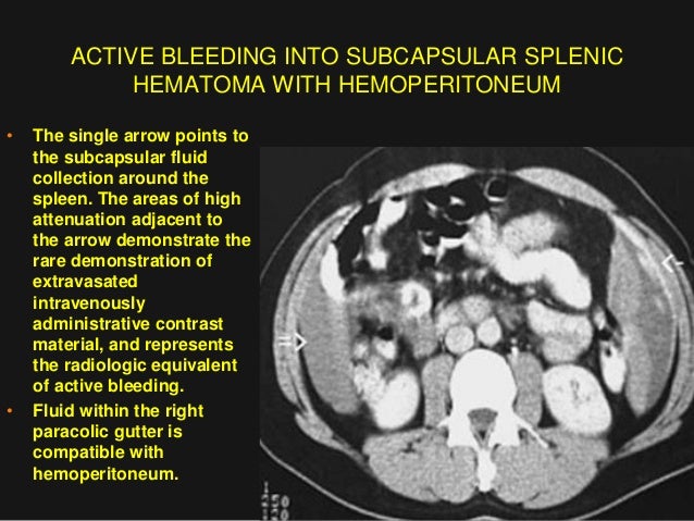
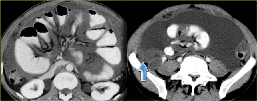
Postoperative computed tomography scan of the abdomen showing extensive hemoperitoneum below the spleen in the left paracolic gutter in front of urinary bladder measuring 12 cm in dimension and a distended rectum measuring 8 5 5.
Paracolic gutter hematoma.
Infected peritoneal fluids get a passageway through these gutters to other compartments of the abdominal cavity.
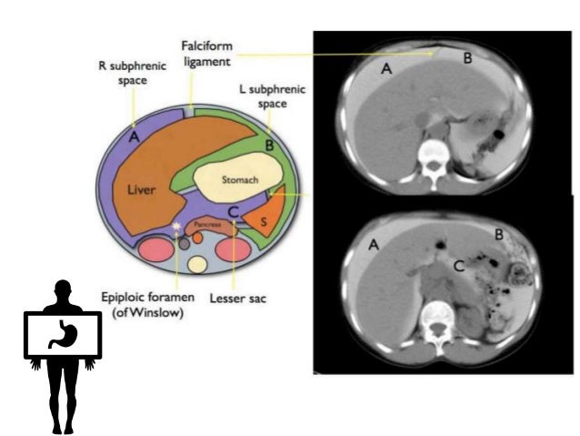
The right and left paracolic gutter are connected to subphrenic spaces proximally and to the pelvic area at the distal end.
In a male patient this is a very uncommon diagnosis.
There is a multi cystic mass extending from the pelvis along the right paracolic gutter to the upper abdomen.
A less obvious medial paracolic gutter may be formed especially on the right side if the colon.
The paracolic spaces gutters are located lateral to the peritoneal reflections of the left and right sides of the colon fig 8a.
Urgent laparotomy revealed an extensive hemoperitoneum.
Ipsilateral psoas hematoma and fat stranding in the right paracolic gutter confirmed rupture of the hemorrhagic cyst from the right native kidney fig.
The right lateral gutter is much larger and allows for greater drainage than the left gutter.
The right and left paracolic gutters are peritoneal recesses on the posterior abdominal wall lying alongside the ascending and descending colon.
Spontaneous abdominal hemorrhage is defined as the presence of intraabdominal hemorrhage from a nontraumatic and noniatrogenic cause.
This is followed by accumulation of ascites in sub hepatic location in 86 cases 86 in morrison s pouch in 85 cases 85 in paracolic gutters in 78 cases 78 in pelvis in 64 cases 64.
Liver or splenic hemorrhage more typically descends peripherally along the paracolic gutters into the pelvis and is not entrapped in interloop spaces.
Paracolic gutters help keep infectious material away from the body s internal organs.
The right paracolic gutter is larger than the left and communicates freely with the right subphrenic space.
The retroperitoneal hematoma measured 13 4 mm diameter and severely compressed the inferior vena cava ivc fig.
Both paracolic gutters run laterally along the back side of the abdominal wall and are situated between the abdominal wall and the outer margin of the colon.
These images look quite similar to images of a pseudomyxoma peritonei which was discussed before.
Common sources of spontaneous abdominal hemorrhage are visceral hepatic splenic renal and adrenal gynecologic coagulopathy related and vascular.
The left medial paracolic gutter.
Thus centrally located triangular areas of high attenuation abdominal fluid should prompt a search for intraperitoneal bowel or mesenteric injury 7 8.
Spontaneous liver haematoma as a result of thrombolytic therapy.
The main paracolic gutter lies lateral to the colon on each side.


